Aim: To evaluate the accuracy of digital impression
by using scan bodies of different geometry and
materials for full arch implant prosthesis.
Settings and Design: This was a systematic review
and meta-analysis following the Preferred Reporting
Items for Systematic Reviews and Meta-Analyses
(PRISMA) guidelines.
Methods and Material: An electronic search
of PubMed (including MEDLINE), EBSCO host
databases, Cochrane library and Google Scholar
search engine for articles published from January
2011 to May 2023 was conducted. The literature
search intended to retrieve all relevant clinical and
in vitro studies about the effect of scan bodies on the
accuracy of digital impression in fully edentulous
arches for full arch implant prosthesis.
Statistical analysis used: Meta-analysis was
conducted in from the reported quantitative data.
Results: A total of 1166 articles were obtained via
electronic search; 8 studies met the inclusion criteria
and were included in this systematic review and were
all in vitro studies. Among the different parameters
described, the scan body material and geometry were
evaluated. Accuracy was measured by evaluating
the linear and angular discrepancies. Among the
8 included studies in this systematic review, only 3
studies were selected for meta-analysis as they were
relatively homogenous in their study design and
outcome variables. Linear discrepancies along X, Y
and Z axis showed a statistically significant difference
between PEEK and Titanium scan bodies (P < 0.05,
pooled mean difference ranging from 0.00 to 0.07)
Conclusions: There is an overall increase in
dimensional accuracy of digital impression recorded
by scan bodies of cylindrical and simpler geometry.
In terms of materials, PEEK scan bodies reported
least discrepancies, thereby deeming to be more
accurate than Titanium scan bodies.
Key words: Accuracy, edentulous, scan bodies, digital impression
Tooth loss results in impairment of masticatory
function, speech, aesthetics, and also affects the
psychology of the patient. Implant-supported
prosthesis, to replace naturally missing teeth, is
one of the most common treatment modalities.1 An
accurate implant impression is an essential pre
requisite for implant restorations, as inaccurate
transfer of the implant position can lead to an ill
fitting prosthesis, which may induce unnecessary
strain on several prosthetic components and may
result in various complications. Moreover, there
is no intervening periodontal ligament at the
implant-bone interface to compensate for any
inaccuracies. The different factors that influence
the implant impression accuracy include the
impression techniques, materials used, and the
number of implants present.2
There are two main conventional implant
supported impression techniques: the direct/
pick-up technique that uses an open-tray; and the
indirect/ transfer technique that uses a closed
tray. The open tray technique is chiefly indicated
when the implants are not oriented parallel
to each other, and can further be subdivided
into splinted and non-splinted techniques. The
closed tray technique is mainly indicated in
case of restricted mouth opening, limited access
areas (posterior region) or in patients with strong
gag reflex.3
Nowadays, digital impressions are widespread
and have revolutionized the field of implantology.
Compared to traditional implant impression
techniques, digital impressions eliminate several
procedures such as dispensing and setting of
impression materials, disinfection, and stone
cast pouring. Also, the simplified workflows not
only improve time efficiency, but also reduces
the possibilities of deformation.4
Digital impressions can be achieved with the help of scan bodies. In the field of implantology,
standardized scan bodies that are inserted
onto the implant instead of impression copings,
have been developed and well established.
This enables computer-aided determination
of the actual implant position using data from
the digital scanner.5 Implant Scan bodies are
precision attachments that are screwed onto the
coronal portion of the implant and reproduces
its position in the digital model. They also assist
on the digital transfer of 3-dimensional position
of dental implants from the patient’s mouth to
computer-aided design (CAD) softwares.6
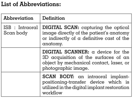
Implant scan body (ISB) characteristics such as
connection type, geometry, dimensions, material
and reusability can play a significant role in
the overall accuracy of the intraoral digital
impression.7 Currently, there are different types
of scan bodies of respective implant system that
are available in the market. These scan bodies
are manufactured as monolithic components
or by a combination of different materials, as
titanium alloy, polyetheretherketone (PEEK),
aluminum alloy, and various resins. Scan bodies
from different manufacturers also differ in their
characteristic geometries as well, such as Flag
shaped, Cylindrical, Tapered, Straight with/
without bevel.7
However, there is limited literature on the effects
of ISB material and geometry on the accuracy
of digital impressions made in fully edentulous
arches. Hence, there is a need of more detailed
investigations on these parameters of scan
body which would be helpful for more accurate
reproduction of the full mouth implant supported
prosthesis. Therefore, the purpose of this
systematic review is to evaluate the accuracy
of digital impression by using scan bodies of
different geometry and material for full arch
implant prosthesis.
This systematic review was conducted according
to the Preferred Reporting Items for Systematic
Reviews and Meta-Analyses (PRISMA)
guidelines8 with prior registration in PROSPERO
(Registration number CRD42023433845). The
focused question was “Will different geometry
and material of scan bodies have an effect
on the accuracy of digital impression for
fully edentulous arches for full arch implant
prostheses?” The PICO i.e., the Population,
Intervention, Comparison, and Outcome format
was used (Table 1). The inclusion criteria were
studies that evaluated the effect of Scan bodies
in fully edentulous arches for full arch prosthesis,
studies on accuracy of digital implant impression
by using scan bodies and articles appearing in
the English dental literature, published after year
2011 till 31st May 2023. The exclusion criteria
were studies wherein the use of scan bodies was
limited to partially edentulous arches. Review
articles, case series and case reports were also
excluded.
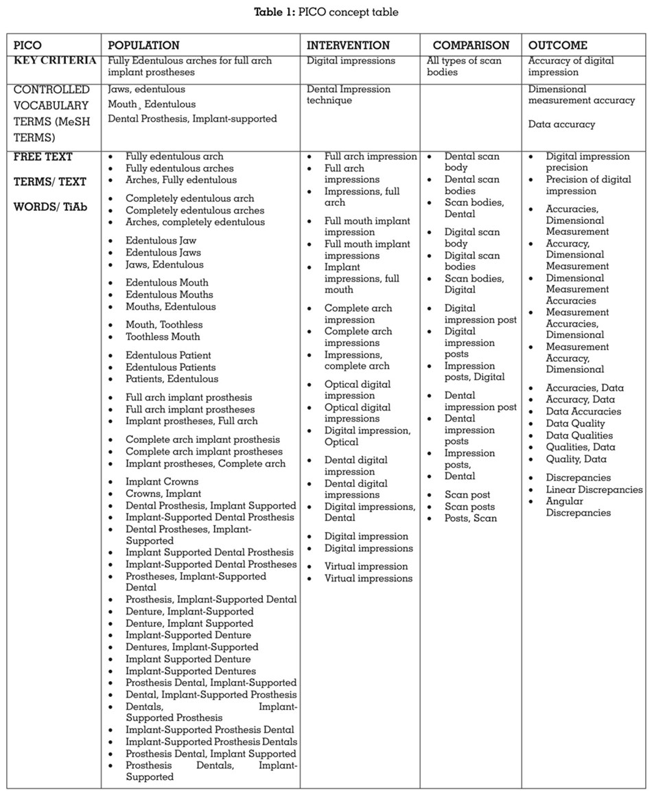
Electronic search of PubMed (including
MEDLINE), Cochrane Central, EBSCO host databases and Google Scholar search engine
for articles published from 1st January 2011 to
31st May 2023 was conducted. The controlled
vocabulary terms (i.e., MeSH terms) and free
text terms were obtained by searching key
concepts in the MeSH database and a thorough
evaluation of related articles, thesaurus,
dictionaries, and entry terms. The terms such as
edentulous jaws, edentulous mouth, edentulous
patients, fully edentulous arches, completely
edentulous arches, full mouth implant
impressions, digital impressions, dental digital
impressions, complete arch impressions, virtual
impressions, dental scan bodies, digital scan
bodies, dimensional measurement accuracy,
data accuracies were combined using suitable
Boolean operators (AND, OR, NOT) (Table 2).
An electronic search was conducted
independently by two reviewers (S.K., A.P.) A
total of 1166 articles were obtained via electronic
search. The articles thus obtained were
evaluated for duplicates. A detailed summary of
data selection has been put forth in the PRISMA
2009 Flow Diagram8 (Figure 1). The study
characteristics of each systematic review were
extracted including study details, search details,
analysis and results/findings by two independent
reviewers (S.K., A.P.) A third reviewer (N.P.S.) was
called in for a final decision if any disagreement
persisted between the two calibrated reviewers.
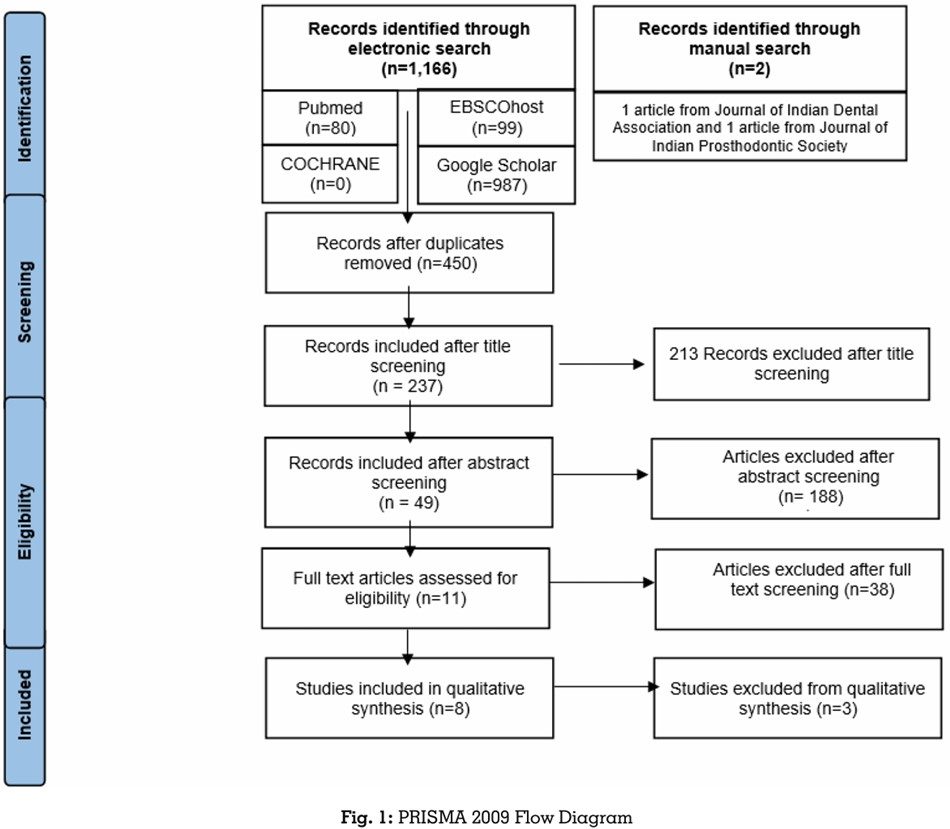
The 1166 articles that were obtained through
the electronic searches were compared
meticulously with respect to the author’s name,
year of publication, title, abstract as well as the
journal name, issue and volume number. The
articles thus obtained after the electronic and
manual searches, were evaluated for duplicates
using the Mendeley Desktop software (v1.19.6).
The 2 articles obtained through the manual
search were added manually using the ‘add entry manually’ feature of Mendeley Desktop
software (v1.19.6). Duplicates were identified
and removed using the software’s “Check for
Duplicates” feature. 716 duplicate articles were
identified and subsequently eliminated leaving
behind 450 articles. Two calibrated reviewers
(S.K., A.P.) independently screened the relevant
titles of the studies found through the electronic
search. Out of 450 articles, 213 articles were
excluded after screening of the title. The articles
thus eliminated were either literature reviews,
scoping reviews, case reports, case series, or
articles on utilization of scan bodies on partially
edentulous arches. Thus, 237 articles were
selected after title screening.
Two calibrated reviewers (S.K., A.P.) now
independently screened the abstracts of the
studies found relevant during the screening of
the titles and a total of 188 articles were further
excluded after abstract screening. The articles
eliminated through abstract screening were
mainly involving different impression materials
and comparing different brands of scan
bodies. 49 articles were included after abstract
screening. Hence, 8 articles were selected after
abstract screening and thus were included in
this systematic review. All the 8 included articles
were in-vitro studies (Supplementary Table 1
and 2).
A third reviewer (N.P.S.) was called in for a
final decision, if any disagreement over article
selection persisted between the two calibrated
reviewers. Inter-reviewer reliability was checked
via Cohen’s kappa coefficient.9 The Cohen’s
kappa coefficient values obtained for title,
abstract and full text screening was 0.65, 0.72
and 0.68 respectively, indicating moderate inter
reviewer agreement for title, abstract and full text
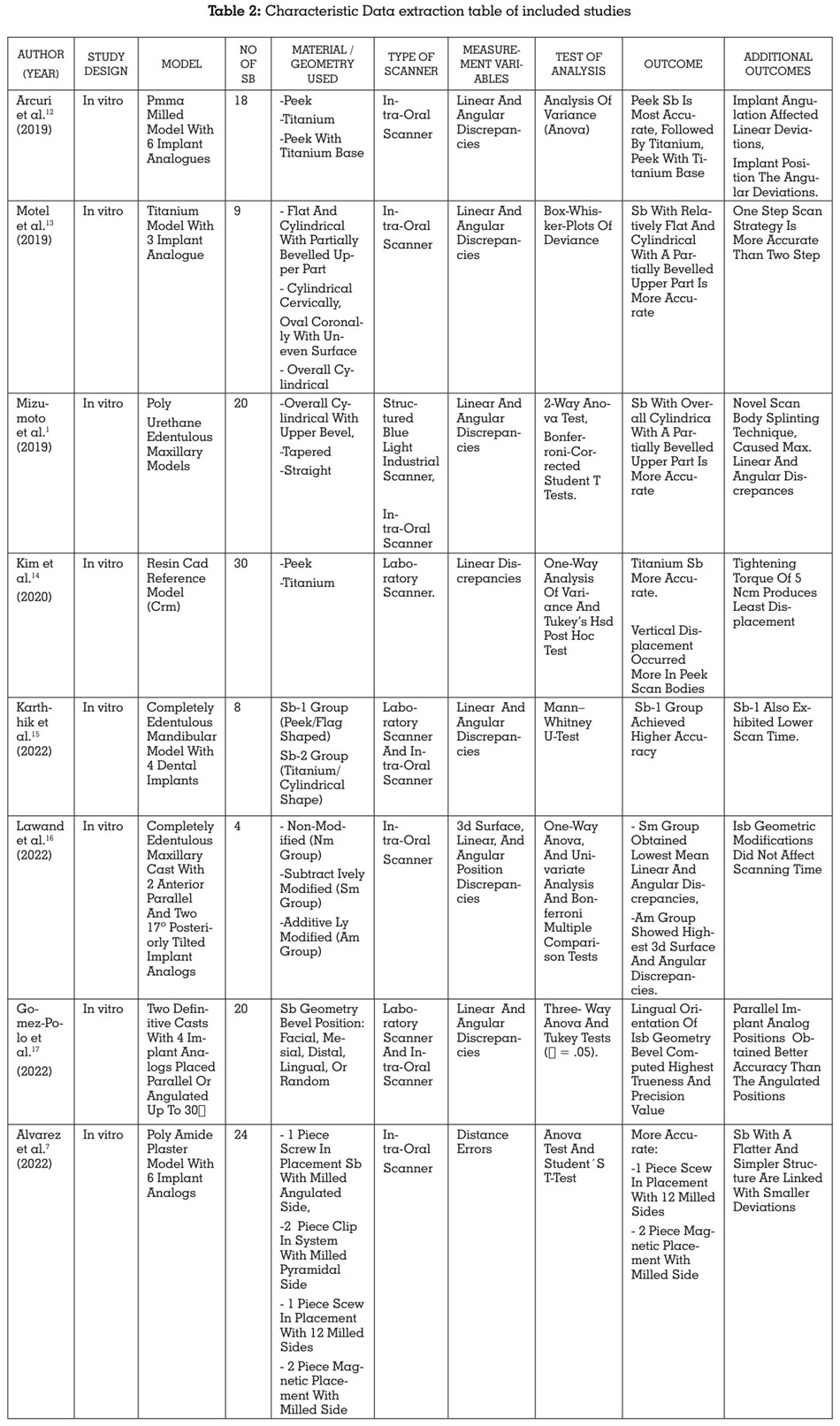
screening. The data was subsequently extracted
from the 8 included studies and recorded in 2
excel data extraction sheets as mentioned in the summary table (Table 2)
1. DIFFERENT SCAN BODY MATERIALS
2. DIFFERENT SCAN BODY GEOMETRIES
The data extracted was entered under the
following headings: Author and Year of
publication, Study design, Study model,
Number of ISB used, Number of scans, Material/
Geometry of Scan body, Type of digital scanner
used, Measurement variables, Test of Analysis,
Outcome and Additional Outcomes
Risk of bias assessment of the included studies was done using the QUIN tool scale10 by two
independent reviewers (S.K., A.P.) This scale is
primarily used to assess the risk of bias of in-vitro
studies. Since all the 8 included studies were in
vitro studies, this scale was considered apt for the
risk of bias evaluation in this systematic review.
The changes made to the scale were validated by
the third reviewer (N.P.S.) In this scale, the items
are scored 0 if not specified, 1 if inadequately
specified or 2 if adequately specified. The results
were then summed to obtain an overall score for
a given in vitro study. The scores thus obtained
were used to grade the in vitro study as high,
medium, or low risk (>70%=low risk of bias,
50% to 70%=medium risk of bias, and <50%=
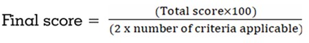
high risk of bias) by using the following formula :
The risk of bias of all the 8 included studies
ranged from 79% to 91%, which falls under the
category of low risk of bias.
Eight studies evaluating the accuracy of digital
impression with different geometry and material
of scan bodies for fully edentulous arches were
included in the systematic review. Five studies
which evaluated the effect of different geometry
of scan bodies on accuracy (Mizumoto et al.,2019;
Motel et al., 2019; Alvarez et al., 2022; Lawand
et al.,2022; Gomez-Polo et al., 2022)1,13,7,16,17
were excluded from meta-analysis due to lack of uniformity in the comparison groups. Three
studies which evaluated the effect of PEEK vs
Titanium material scan body on the accuracy of
digital impression (Arcuri et al., 2019; Kim et al.,
2020; Karthhik et al., 2022)12,14,15 were included
for meta-analysis.
The Review Manager software (Version 5.4.1)
was used to perform meta-analysis. Mean
values and standard deviations for linear
discrepancies were included for the analysis.
The linear discrepancies were measured along
three axis- X, Y and Z axis. The primary outcome
measures the accuracy of digital impression,
and was evaluated by measuring the linear
deviations along X, Y and Z axis. More the linear
deviations present, lesser is the accuracy. The
data was tabulated under the headings of study
name, group, and effect size. The effect size was
calculated on the continuous raw data entered
for mean, standard deviation and sample size.
95% confidence interval for each effect size was
also computed. The heterogeneity of effects was
assessed by the Higgin’s I2 test.11
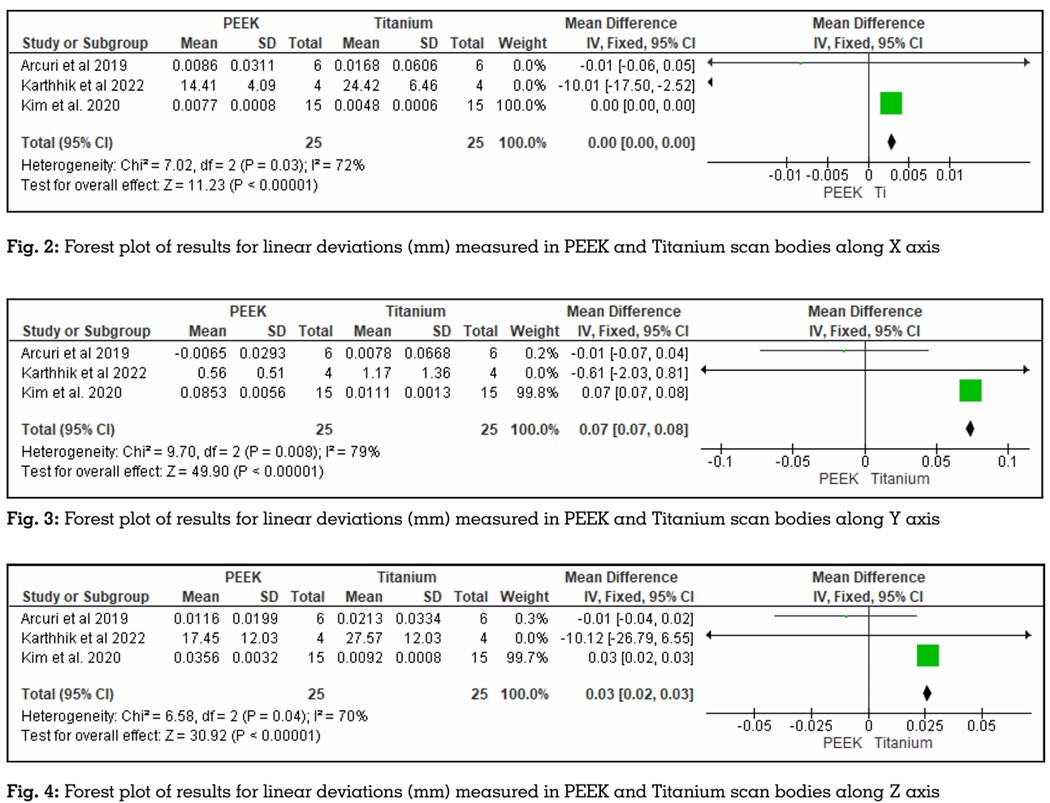
A statistically significant difference was observed
between the two materials of scan body along
X axis (P < 0.05, pooled mean difference =
0.00 CI = 95%), along Y axis (P < 0.05, pooled mean difference = 0.07 CI = 95%), and along
Z axis (P < 0.05, pooled mean difference =
0.03 CI = 95%) as stated in the forest plot. The
results of the meta-analysis for linear marginal
discrepancy showed minimum discrepancy in
PEEK and maximum discrepancy in Titanium
scan body along X, Y and Z axis (Figures 2, 3, 4
respectively)
This systematic review analyzed the effect of the
various characteristics of ISBs on the accuracy
of implant impression, and is chiefly based on
in vitro studies. Among the parameters assessed in this study were the scan body material and
geometry.
The ISB material is a crucial factor that has to be
scrutinized, as it can have a significant impact
on the biocompatibility and scanning accuracy
which depends on surface reflections and in turn
can influence the number of stitching points in
order to attain the desired results.12 Different
materials which are used include polymers
such as polyetheretherketone (PEEK), titanium
alloys, aluminum alloys, and resin materials.
PEEK and Titanium are the most commonly used
for ISB fabrication. PEEK is high-performance
thermoplastic polymer with excellent physical and mechanical properties, chemical stability
and low weight. It is often used in ISBs because
it can easily be scanned compared to other
materials and it does not cause any surface
reflections which could cause hinderance in
intraoral scanning.12
However, the selection of polymeric materials
has recently come into question, as repeated
sterilization, clamping forces, and even chewing
forces can deform and abrade the polymeric
materials. This is the reason why PEEK ISBs
are suggested only for single usage. Ti-alloy
components are also commonly selected for
the fabrication of ISB. It represents excellent
biocompatibility and is resistant to deformation
on repeated sterilization.13 Hashemi et al
evaluated the effect of repeated use of PEEK and
Titanium Scan Bodies on the transfer accuracy
of implant position and demonstrated that the
inter-implant distance variations were more
in titanium as compared to PEEK scan bodies.
The results further indicated that titanium scan
bodies had lesser dimensional changes as
compared to PEEK scan bodies after repeated
use.19
Nevertheless, there were inconsistencies
between the different studies about which is the
preferable material for ISB. In a study given by
Arcuri et al, PEEK ISBs showed optimal results on
both linear and angular measurements, which
was followed by Titanium. PEEK with titanium
base showed least accuracy because of its bi
component configuration.12 In a study given by
Karthhik et al, PEEK SB material showed more
scanning accuracy as it reduced the problem
of light reflectance that can occur in the metal
alloy.15 PEEK showed optimal results on both the
studies however this was in disagreement with
another study by Kim et al, which presented better
trueness and stability of the Ti-ISB compared to
PEEK ISBs.14
Another characteristic feature that was assessed
in the included studies was the ISB geometry.
A relatively flat, more simply constructed scan
body resulted in significantly smaller deviations
within the digital impression. In two studies
ISB with overall cylindrical geometry with a
partially beveled upper part showed better
scanning trueness in distance deviation.1,6 The
current study found out that worst results were
seen in scan bodies which had a very complex
anatomy with irregular surface. This finding
agrees with the study published by Kurz et al,
which shows that more intricate the scan body
surface, as in sharp edges, more are the errors
registered.20 The ISB geometry bevel also has a
significant effect on the scanning accuracy and
the lingual placement of the bevels is proposed
for best results.17 An interesting finding is that
the scanning trueness of ISBs was improved by
subtractive modifications in design, whereas it
was reduced by additive alterations.18 Surfaces
which are more challenging to scan include
steep, sharp, deep undercuts, angled or
overcrowded surfaces.
The position and location of implant and ISB
has impact on the scanning trueness in distance
deviations. If the most distal implant was tilted
mesially, there would be better trueness in
scanning accuracy. The extent of edentulism
is another important factor; Free-end partial
edentulism (Kennedy I, II) results in higher
deviations compared to Kennedy III, IV. This
is can be possibly attributed to inter-implant
space which is limited in Kennedy I and II
cases.21 In this systematic review, majority of
studies had linear and angular discrepancies
as measurement variables. In terms of material,
PEEK showed least discrepancies, followed by
Titanium followed by PEEK with Titanium base.
Considering the geometrical aspect of ISB, Scan
bodies with cylindrical and simple configuration
showed least discrepancies.
The QUIN Tool has been used here based on the
in-vitro study design of the included studies to
identify the risk of bias of the individual studies.10
Three out of 8 included studies seemed to be
relatively homogenous in their study design and
outcome variables. Hence, a quantitative analysis
by means of a meta-analysis was planned.
Meta-analysis is a systematic procedure that
is used for assessing and combining statistical
information based on results of available
independent studies regarding the same topic.
The results of the quantitative analysis have
been provided in the form of forest plots for easy
visualization. The heterogeneity of the primary
studies has been evaluated using the Higgins’s
I2 test.11 Heterogeneity refers to differences in
results between primary studies that are greater
than expected by chance alone. The results of the
meta-analysis for linear marginal discrepancy
showed minimum discrepancy in PEEK and
maximum discrepancy in Titanium scan body
along all three planes.
After evaluating different studies in the current
systematic review, PEEK biomaterial scan bodies
do show a promising outcome for better accuracy
for full arch implant supported prosthesis as
compared to that of Titanium scan bodies. Also,
when it comes to the geometry of scan bodies, it
can be said that scan bodies with least complex
geometry will show minimal errors during
scanning, especially in full arch cases. Hence,
clinicians will have a better understanding
in selection of scan bodies when it comes to
scanning of fully edentulous arches.
Limitations of this systematic review were that
all the included articles were in vitro in their
study design, as there were nearly no clinical
studies in the available literature. The search for
this study was also limited to articles published
in the English language only. Other ISB
parameters like location of ISB, wear, inclination
of dental implants and ISB dimensions were not included in this systematic review as it would
contribute more to the heterogeneity of the
current systematic review. Hence, the results of
this systematic review should be applied with
caution to the clinical scenario and more in vivo
randomized controlled trials should be carried
out to support the current evidence.
Although intraoral scan bodies and their characteristics vary widely, they significantly influence implant impression accuracy. The majority of studies agree that, among the various characteristics, the material, and the geometrical design affect the impression accuracy significantly.
These conclusions enable the clinician in
proper decision making to choose the PEEK
scan bodies with simple geometry whenever
possible for digital impressions of their full arch
implant cases. However, more clinical studies
are necessary for safer conclusions, since the
available scientific evidence is not yet conclusive
about the optimal Intraoral Scan Body.