Purpose: To evaluate the effect of adding 1wt% of
Al2O3, SiO2, and ZrO2
nanoparticles to heat cure
acrylic resins on their flexural strength.
Materials and Methods: PMMA is the widely
accepted denture base material but has got the
disadvantage of low mechanical strength. So
the study evaluates whether reinforcement with
nanoparticles improves its flexural strength. A total
of 88 specimens are prepared in 4 groups (Control,
1wt% Al2O3, 1wt% SiO2, 1wt% ZrO2) and 3 subgroups
(5 nm & 15 nm of all Nano groups) (n =22/group). The
PMMA powder and nanoparticles are mixed with
each other by means of ball milling and is invested
in type III dental stone for processing. Flexural
strength is measured via three-point bending
tests. Subsequently, SEM analysis is performed for
specimens from each group to ensure homogenous
distribution.
Results: The flexural strength of
polymethylmethacrylate (PMMA) after adding 1wt%
of aluminium trioxide 15nm significantly increased
(p ˂ 0.05) followed by 1wt% of silicon dioxide 15nm.
The scanning electron microscopy analysis revealed that the particles were homogeneously dispersed
in PMMA matrix.
Conclusion: The mechanical properties of heat cure
PMMA can be increased by addition of nanoparticles
to PMMA powder. 1wt% of aluminum trioxide-15nm
diameter has significantly improved the flexural
strength of PMMA.
Key words: nanoparticles, Flexural strength, Aluminum trioxide, Zirconium dioxide, Silicon dioxide.
Denture base is defined as the part of a denture
that rests on foundation tissue to which the teeth
are attached. The denture base material is
defined as any substance of which the denture
can be made. -GPT9
Poly (methyl methacrylate), is widely used in
rehabilitation of edentulous patients because of
its satisfactory esthetics, ease of use, low cost,
light weight and stability in the mouth5,8. Many
different materials such as bone, wood, ivory, vulcanized rubber, polystyrene, light activated
UDMA are used but PMMA remains preferred.
But the material has the disadvantage of low
mechanical properties such as low flexural
strength and impact strength resulting in fracture
of the denture when subjected to occlusal flexural
strength of standard denture base polymers
prescribed is 65 MPa by the ISO 1567.
Nanoparticles are effective reinforcing materials
and used as fillers for polymeric materials
to improve their mechanical properties. They
are characterized by their smaller size, larger
specific surface area, and also strong interfacial
interaction with organic polymers. The dimension
of the material is in nanometer, which leads to
unique properties. In the current study 1wt% of Al2O3, SiO2, & ZrO2 of diameter 5nm and 15nm
are added to improve the mechanical properties.
The purpose of the study is to evaluate the flexural strength of heat cure PMMA on adding 1wt% of Al2O3, SiO2, & ZrO2
of diameter 5nm and 15nm.
Polymer is a macromolecule composed with
repeated unit. They are formed by condensation
polymerization or addition polymerization.
Acrylic resins are polymer esters of methacrylic
acids. PMMA is the combination of methyl
methacrylate with chemical formula (C5H8O2) n.
PMMA is a linear thermoplastic polymer which
lack of methyl group on its backbone carbon
chain.
PMMA is commonly used denture base material. The PMMA employed in the current study is
IVOCLAR Vivadent SR triplex hot pink heat cure
veined denture base material. The powder liquid
ratio is followed according to the manufacturer’s
instruction as 20 ml for 43.8 gm. of powder by
means of measuring jar and graduated cylinder.
The specimens of the dimension 65mm in length,
10mm wide and 3 mm thickness are fabricated as per the ISO 1567 standard, by using templates.
In the present study, three different nanoparticles
were added in ratio of weight 1 wt% nanoparticles (Al2O3, ZrO2, SiO2) to the heat cure acrylic
resin. 1wt% nanoparticles (Al2O3, ZrO2, SiO2) has
been used so as to prevent agglomeration and
to ensure homogenous distribution without adversely affecting the mechanical properties.
These were categorized into the following
4 groups pure PMMA – Group I, PMMA with 1wt%
Al2O3
-Group II, PMMA with 1wt% SiO2
– Group
III, PMMA with 1wt% ZrO2
– Group IV and 3 sub-groups PMMA with 5nm 1wt% Al2O3
(Sub Group
II a) & 15 nm 1wt% Al2O3
(Sub Group II b), PMMA
with 5nm 1wt% SiO2 (Sub Group III a) & 15nm
1wt% SiO2
(Sub Group III b), PMMA with 5nm
1wt% ZrO2
(Sub Group IV a) & 15 nm 1wt% ZrO2
(Sub Group IV b).
The investment is made using type III gypsum
product. The PMMA powder and Nano particles
are mixed by means of ball milling machine
(RETSCH PM 100). The speed is set at 350 rpm
for one hour with four steel balls of diameter
10mm with an interval at 30 minutes.
The PMMA powder (IVOCLAR Vivadent SR
Triplex Hot) along with the nanoparticles (Matrix
Nano) is mixed with the liquid (monomer) and
left for 10 minutes and then packed into the flask.
The flask is approximated and kept for curing in
the curing unit (DELTA – Tempsen Polybath) for
two hours and then boiled for 1 hour. The flasks
were allowed to cool for 30 minutes and placed
under running water for 20 minutes to ensure
complete cooling. Then the specimens were
deflasked, trimmed and polished using 200, 400
and 600 grit sand paper and the specimens are
immersed in distilled water. The samples are
then subjected to three-point bending test using
universal testing machine (INSTRON 3345 –
Bluehill 3). After being subjected to three-point
bending tests the specimens are analyzed under
scanning electron microscope (CAREL ZEISS EVO18) to ensure homogenous distribution of
nanoparticles.
The data was expressed in mean and standard
deviation. Statistical Package for Social Sciences (SPSS 20.0) version used for analysis. Oneway ANOVA (Post hoc) followed by sh used for
analysis. p value less than (p<0.05) considered
statistically significant at 95% confidence
interval.
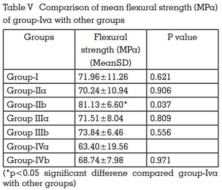
The values of control group exhibit a maximum
flexural strength value of 94.3 MPa and a
minimum of 55.8 MPa with an average mean of
71.96 ± 11.26 MPa.
The flexural strength of heat cure PMMA reinforced with 1wt% of 15 nm aluminum trioxide
(81.13 ± 6.60 MPa) (table III) exceeded those of
other groups followed by 1wt% of 15nm Silicon
dioxide (73.84 ± 6.46 MPa) (table III) and the
group reinforced with 1wt% of 5 nm Zirconium
dioxide (63.40 ± 19.56 MPa) (table III) had the
least strength. The values of 15nm Al2O3
and
15nm SiO2
were found to be statistically significantly higher than those of the control group (p
< 0.05).
PMMA is commonly used material for denture
base because of its ease of use, polishability
and light weight. But it has got the disadvantage
of low flexural strength and impact strength resulting in the fracture of denture. Reinforcement
with nanoparticles is expected to improve the
mechanical properties.
The present study evaluated the flexural strength
of heat cure PMMA reinforced with different nano
particles and sizes. Different nanoparticles, such
as aluminum trioxide, silicon dioxide, zirconium
dioxide of 5nm and 15nm diameter were used.
According to the study conducted by Karci et
al34 particles of size as low as 15 nm produced
better results so in the current study comparison
between 5nm and 15 nm were done to test the efficacy. The mean flexural strength of the control
group obtained is -GROUP I- 71.96 ± 11.26 MPa.
The mean flexural strength of other groups when
compared to the control group clearly states that
group IIb (81.13 ± 6.60 MPa) (table III) has the
highest values followed by Group IIIb (73.84 ±
6.46 MPa) (table III). Group IVa has got the lowest strength (63.40 ± 19.56 MPa) (Table III)
The minimum value of flexural strength of standard denture base polymers prescribed is 65
MPa by the ISO 1567. The values (table III) of
Group IIa (70.24 ± 10.94 MPa), IIb (81.13 ± 6.60
MPa), IIIa (71.51 ± 8.04 MPa), IIIb (73.84 ± 6.46
MPa), IVb (68.74 ±7.98 MPa) and control - group
I (71.96 ± 11.26 MPa) satisfies this requirement.
The values of group IVa - zirconium dioxide 5nm
(63.40 ± 19.56 MPa) is less than that of the ISO
1567 requirement. On comparing mean flexural strength of aluminum trioxide 5nm (70.24 ±
10.94 MPa), silicon dioxide 5nm (71.51 ±8.04
MPa) and Zirconium Dioxide 5nm (63.40 ±19.56
MPa) the silicon dioxide 5nm had the highest
mean flexural strength. On comparing mean
flexural strength of aluminum trioxide 15nm
(81.13 ± 6.60 MPa), with silicon dioxide 15nm
(73.84 ± 6.46 MPa) and Zirconium Dioxide 15nm
(68.74 ± 7.98 MPa) the aluminum trioxide 15nm
(81.13 ± 6.60 MPa) had the highest mean flexural strength (Table IV).
On comparing mean flexural strength of aluminum trioxide 5nm (70.24 ± 10.94 MPa) and 15nm
(81.13 ± 6.60 MPa) no significant difference was
observed. The p value is 0.502 (P>0.05).On comparing mean flexural strength of Silicon Dioxide
5nm (71.51 ± 8.04 MPa) and 15nm (73.84 ± 6.46
MPa) no significant difference was observed. The
p value is 1.00 (P>0.05). On comparing mean flexural strength of Zirconium Dioxide 5nm (63.40 ±
19.56 MPa) and 15nm (68.74 ± 7.98 MPa) no significant difference were observed. The p value is
0.971 (P>0.05). On comparing the mean flexural
strength of Group IIa with other groups no significant difference were observed (p>0.05). On
comparing the mean flexural strength of Group
IIb and Group IVb with other groups significant
difference were observed (p<0.05). On comparing the mean flexural strength of Group IIIa and
Group IIIb with other groups no significant difference were observed (p>0.05). On comparing
the mean flexural strength of Group IVa with other groups significant difference were observed
(p<0.05) (Table V).
Multiple comparison of mean flexural strength
(MPa) between the groups states there is significant difference on comparing Group III a with
Group IVa. (p<0.05). In accordance with the
study conducted by Unal et al39 the shape and
size of the filler particles, distribution in the polymer matrix, and connection to the matrix play a
very important role, and also the size of metal
oxides should be sufficiently low for homogenous mixtures. This prevents a heterogeneous
mixture, and nanoparticles will fill in the cracks
between polymer matrix, thereby preventing the
movement of the chain. In addition to that, the
percentage of the nanoparticles should be kept
low so that the particles will be embedded in the
resin without agglomeration. Balos et al23 also
concluded that low concentration provides better properties.
According to the study conducted by Mahroo
et al49 2.5 wt% of aluminum trioxide with grain
size of 3 micrometer significantly increased the
flexural strength. The addition of 5 wt% Al2O3
powder caused a 5.82% reduction in flexural
strength. Possible explanations for this decrease
in strength could be a decrease in cross-section
of the load-bearing matrix of polymer; stress
concentration due to too many filler particles;
mode of crack propagation through the specimen because of increased amount of fillers; void
formation from the entrapped air and moisture;
incomplete wetting of fillers by resin; and acts as
an interfering factor in the integrity of the polymer matrix. In this study, 1wt% has been used so
as to prevent agglomeration and to ensure homogenous distribution without adversely affecting the mechanical properties.
According to the study conducted by Neveen et
al11 High Impact acrylic resins reinforced with
5% and 15% ZrO2
showed that it increased flexural strength and the flexural strength was proportional to the concentrations. In contrast to this, Ihab and Moudhaffar28 compared the flexural strength after adding ZrO2
nanoparticles
and stated a statistically significant reduction of
more than 5%. So addition of nanoparticles can
influence the flexural strength both in a positive
as well as negative aspect.
According to the study conducted by Reem
Abualsaud, the Nano SiO2
particles increased
the flexural strength. Additionally silane treatment of the nanoparticles enabled stronger
bonds with the matrix.
In the current study highest values are obtained by using 15nm Aluminum trioxide
(81.13±6.60 MPa) followed by 15nm silicon dioxide (73.84±6.46MPa) (table III). The p value
is significant for the 15nm 1wt% Al2O3
(p<0.05).
The zirconium dioxide 5 nm had the lowest value (63.40±19.56 MPa) with a significant p value
(p<0.05).
On comparing the diameter among the same
groups no statistically significant results were
obtained (p>0.05) but still 15nm sub-groups
of all the three major groups had better results
when compared to the 5nm sub-groups (Table
I and Table II). In the study conducted by Karci et al34 the specimens after testing for flexural strength are then subjected to SEM analysis
to ensure homogenous distribution. Similarly in
the present study, specimens after three point
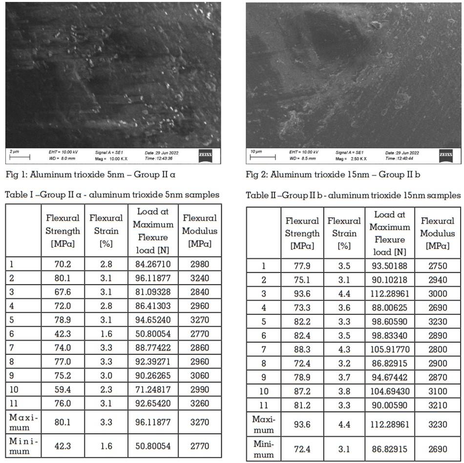
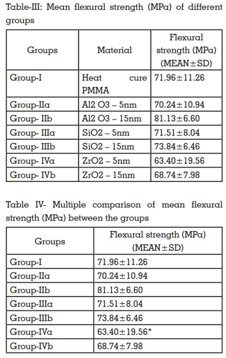
bending test were analysed under SEM for ensuring homogenous distribution. (Fig. 1 and Fig.
2). This homogenous distribution as stated by
Karci et al34 has promoted the mechanical properties of the material. A few studies recommend
using coupling agents to prevent the agglomeration of particles. They have also stated that
the well-distributed particles are only capable of
better stress transfer and reinforcement. The null
hypothesis was rejected, as the flexural strength
has changed after the addition of nanoparticles.
The flexural strength of heat cure PMMA reinforced with 1wt% of 15 nm aluminum trioxide exceeded those of other groups followed by 1wt%
of 15nm Silicon dioxide and the group reinforced
with 1wt% of 5 nm Zirconium dioxide had the
least strength.
Variations in the particle size were found to be
effective in improving the strength. According
to the study conducted by Karci et al34 the nano
particles of diameter as low as 15 nm were yielding a good improvement. In the current study
comparison between 5nm and 15nm was done
to evaluate whether 5nm diameter particles provided better results. The results do not show statistically significant results but 15nm groups had
better results compared to the 5nm sub groups.
In the present study, the saliva was not considered since this study was an in-vitro study,
whether it might affect the results clinically is not
evident. Furthermore, the leaching of particles
might vary the biological properties of heat activated denture base material. This has proven to
be a major limitation of the study and thence an
in-vivo study can be considered in the near future. The optical properties of aluminum trioxide
were not that satisfactory as it caused greyish
discoloration. Henceforth, efforts have to be undertaken for improving the optical properties of
aluminum trioxide nanoparticles.
Reinforcing PMMA with the nanoparticles such as aluminum trioxide, silicon dioxide and zirconium dioxide has proved to be beneficial in improving the mechanical properties. The flexural strength has been increased on addition of these particles. Adding nanoparticles can adversely affect the mechanical properties also. It is observed that Lower concentrations of nanoparticles prevents agglomeration and have better results. Concentration as low as 1wt% has yielded positive results. The Particle size also plays a vital role in ensuring reinforcement. Particle size of about 15nm has yielded better results. The following conclusions can be drawn from this study