JPID - Vol 07 - Issue 02

A NOVEL FABRICATION TECHNIQUE OF PROSTHETIC HOLLOW VAGINAL STENT FOR A CASE OF MAYER–ROKITANSKY– KUSTER–HAUSER SYNDROME

* Teena Wilson, ** Vivek V Nair, ***Harshakumar K, **R Ravichandran
*Postgraduate Student, **Professor, ***Professor & Head of the Department, Department of Prosthodontics, Government Dental College, Thiruvananthapuram - 695 011 | Corresponding Author: Dr. Teena Wilson, E Mail: teenacapricon23@gmail.com

Abstract:

Vaginal stents are most widely employed in the management of maintaining the patency of neovagina in patients with Mayer-Rokitansky-Küster-Hauser (MRKH) syndrome. Although several methods are available for the fabrication of vaginal stents, hardly any techniques are described in the literature to date to make the vaginal stent prosthesis hollow. This article presents a novel simple and cost-effective technique for fabricating custom hollow vaginal stent prostheses for patients with MKRH syndrome. This dental technique offers a convenient removable treatment option for maintaining the patency of the neovagina and is a very straightforward, reasonably simple, and cost-effective method for making vaginal stents.

Key words: Mullerian aplasia, stents, vagina

Introduction

Mayer-Rokitansky-Küster-Hauser (MRKH) syndrome is a congenital disorder in women characterized by agenesis or aplasia of the uterus and upper part of the vagina1–3. The management of vaginal agenesis includes the creation of a neovagina either by surgical or non-surgical approaches4 . Vaginal dilation created by non-surgical methods is considered the first line of management before the commencement of any surgical intervention1. Frank’s method and Ingram’s method are the two most common non-invasive vaginal dilation methods4. The most commonly used method is Frank’s method where progressive dilators are manually placed on the vaginal apex1. Vaginal dilators are also used postoperatively to maintain the width and depth of the neovagina, prevent neovagianl contraction or shrinkage, and serve as a hemostat as well5. Maxillofacial prostheses and the materials used in their construction have today provided prosthodontists with a wide range of options to serve humanity and assist our colleagues in the fields of medicine, surgery, and allied specialties. Prosthetic materials such as acrylic or acrylic stents lined with silicones are widely used for the fabrication of vaginal dilators5–7”. These dilators can be made hollow in order to reduce the weight of the prosthesis thereby providing patient comfort. This article describes a novel simple technique for fabricating custom hollow acrylic surgical stent for patients with Mayer-Rokitansky-Kuster-Hauser (MRKH) syndrome. The patient’s privacy was not violated and informed consent for publication was obtained from the patient.
Technique

  1. Determine the dimensions of the vaginal stent to be fabricated based on the magnetic resonance imaging (MRI) report obtained from the gynaecologist.
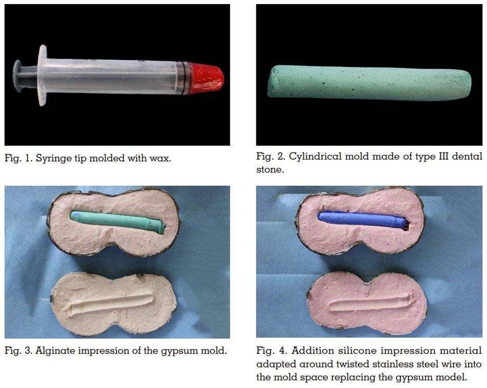
  2. Fabricate the vaginal stent cylindrical mold with a diameter 3mm less than the planned diameter. A 10 ml syringe tip moulded with wax as per the dimensions is selected. (Fig.1)
  3. Make an alginate impression (Zhermack Hydrogum 5 Dental Alginate) on this wax cylinder and pour the mould in type III dental stone (Zhermack Elite Model Type 3 Dental Stone). (Fig.2)


  4. Alginate impression of the same gypsum model is made in a modified brass flask and close tightly with a metal clamp (Fig.3). Replace the gypsum model with polyvinyl siloxane putty consistancy impression material (GC Flexceed putty) adapted around a twisted stainless-steel wire with its two ends projecting out perpendicularly through one end of the putty into the mold space. (Fig.4)
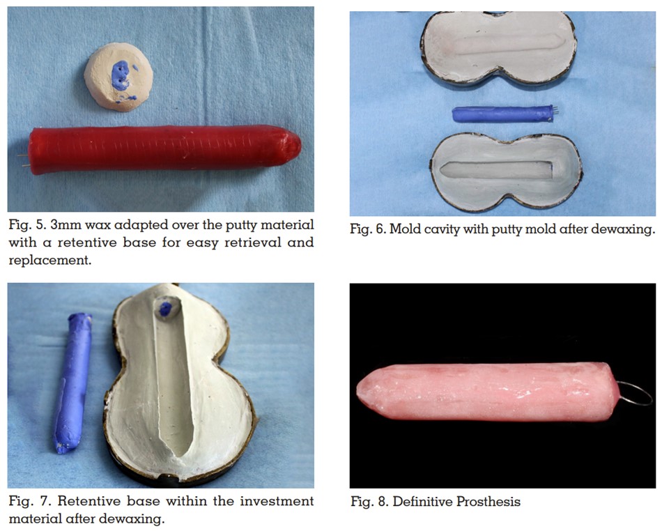
  5. Adapt wax around the putty mold at 3mm thickness so that the predetermined dimensions and shape of the vaginal stent is achieved. A retentive base is also fabricated in putty surrounded with dental plaster inorder to facilitate for easy retreival and replacement of the putty mold during dewaxing and packing procedure. (Fig.5)
  6. Invest the wax pattern in a dental flask. Eliminate the wax by putting the flask in boiling water for 5-7 minutes once the investment material is set. (Fig.6)


  7. Eliminate all the wax residues and detach the putty from the retentive base within the mold space. (Figure.7). Apply cold mold seal all over the mold cavity. Pack an adequate quantity of heat activated acrylic resin into the mold cavity on both halves of the flask. Reattach the putty mold in proper position within the retentive base so that it gets interposed between the two halves. Do trial closure until excess acrylic gets squeezed out around the edges.
  8. Process the stent in heat-activated acrylic resin (DPI Heat cure; Dental Products of India Ltd) by using compression moulding technique. After processing, remove the putty material by pulling the stainless wire to make it hollow in order to reduce the weight of the prosthesis thereby providing patient comfort.. The open end of the acrylic stent is closed with auto polymerizing acrylic resin (DPI RR Cold cure; Dental Products of India Ltd). Finish and polish the vaginal stent to get a smooth surface free of bubbles and sharp projections. (Fig.8)

Discussion

A comprehensive literature review has been conducted by Callens et al. regarding the management of vaginal agenesis and justified vaginal dilators as the first line of treatment8. Vaginal dilators are indicated as a non-surgical intervention method only when the vaginal dimple is deep enough (2–4 cm)5. Postoperatively, these vaginal stents are used to preserve vaginal width and depth, as well as to stop neovaginal stricture and contraction5. Customized vaginal stents or dilators offer great advantages compared to prefabricated ones as it is economical, posses adequate strength and durability9. The hollowed design reduces the weight of the prosthesis making it more comfortable for the patient. Moreover, the heat activated acrylic resin material eliminates the possibility of fungal infections as reported with silicone materials5. The putty mold employed in this technique for hollowing the prosthesis can be effortlessly removed if fabricated without any undercuts. A uniform and adequate thickness of material can be ensured all around the hollow cavity with this technique. Low compliance from the patient side, the time required to achieve satisfactory result and initial discomfort are the possible limitations.

Conclusion

The technique for fabrication of a custom-made hollow acrylic vaginal stent for patients with Mayer-Rokitansky-Küster-Hauser (MRKH) syndrome offers a convenient removable treatment option for maintaining the patency of the neovagina and is a very straightforward, reasonably simple, cost-effective method for making vaginal stents.

REFERENCES

  1. Bombard DS, Mousa SA. Mayer–Rokitansky–Kuster– Hauser syndrome: complications, diagnosis and possible treatment options: a review. Gynecological Endocrinology 2014; 30: 618–623.
  2. Tn E, Ml P, Rl B. Vaginal malformations. Am J Obstet Gynecol 1981; 141: 910–920.
  3. Londra L, Chuong FS, Kolp L. Mayer-Rokitansky-Kuster-Hauser syndrome: a review. International Journal of Women’s Health 2015; 7: 865–870.
  4. Herlin MK, Petersen MB, Brännström M. Mayer-Rokitansky-Küster-Hauser (MRKH) syndrome: a comprehensive update. Orphanet Journal of Rare Diseases 2020; 15: 214.
  5. Kamalakannan J, Murthy V, Kularashmi BS, et al. Customized Silicone Vaginal Stent. J Obstet Gynecol India 2015; 65: 281–283.
  6. Belkhode VM, Nimonkar SV. Hollow vaginal stent for a case of Mayer-Rokitansky-Kuster-Hauser syndrome: a case report. Pan Afr Med J; 38. Epub ahead of print 2021. DOI: 10.11604/pamj.2021.38.343.28837.
  7. Rathee M, Boora P, Kundu R. Custom fabricated acrylic vaginal stent as an adjunct to surgical creation of neovagina for a young female with isolated vaginal agenesis. J Hum Reprod Sci 2014; 7: 272–275.
  8. Update on surgical and non-surgical treatments for vaginal hypoplasia | Human Reproduction Update | Oxford Academic, https://academic.oup.com/humupd/ article/20/5/775/2952651 (accessed 17 March 2023).
  9. Rathee M, Singhal SR, Malik S, et al. Resilient Customized Hollow Vaginal Stent for the Treatment of Vaginal Agenesis in Mayer–Rokitansky–Kuster–Hauser Syndrome. Nigerian Journal of Surgery 2020; 26: 88–91.

JPID – The journal of Prosthetic and Implant Dentistry / Volume 7 Issue 2 / Jan–Apr 2024

Copyright © 2017-25 Indian Prosthodontic Society Kerala State Branch | IPSKERALA.COM | Powered by Dentaura.Com